Early diagnosis of skin cancer is the best tool we have for efficient treatment
At my SKIN SPECIFIC skin cancer clinics, I offer the full range of specialist assessment treatment options:
- Spot checks
- Full body assessment
- Dermatascopic skin assessment
- Shave and punch biopsies
- Liquid nitrogen treatments
- Topical skin treatments with Aldara or Efudix creams
- Surgical excision, including flaps and grafts
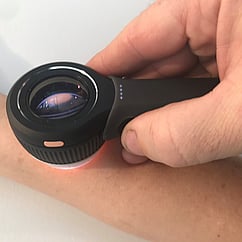
I use dermoscopy for the diagnosis of skin cancers. This is a polarised microscopy system that greatly improves diagnostic accuracy over the naked eye. You can read and see all about the benefits of dermoscopy below.
I also use DermEngine, an advanced software program for capturing, analysing and storing high-resolution photographic images of skin lesions. This is a brilliant tool to monitor and analyse any changes in lesions over time, and an added benefit is that patients can access their own reports.
What happens when you book a skin check?
It’s as easy as walking into your GPs surgery. Having a skin cancer specialist in a general practice setting ensures easy access to this kind of specialised care. We see cases of skin cancer on a daily basis, and we have the specialist training and the necessary experience to ensure excellent clinical management. And best of all, its quick and easy to access. Contact my clinics directly for a booking HERE
How we diagnose – the benefits of dermoscopy
Dermoscopy enables you to see a well-lit, polarised and magnified image of a skin lesion. This combination of magnification and polarised light allows a doctor to look thorough layers of skin, and view sub-surface structures.
This increased accuracy means that skin cancer and pre-cancerous changes are picked up early, and at a very high rate. It also significantly reduces the number of non-cancerous lesions that are removed. This minimises unnecessary treatment and surgery.
The dermatoscope is an amazing tool. Have a look at my gallery of dermascopic images HERE
Types of skin cancer
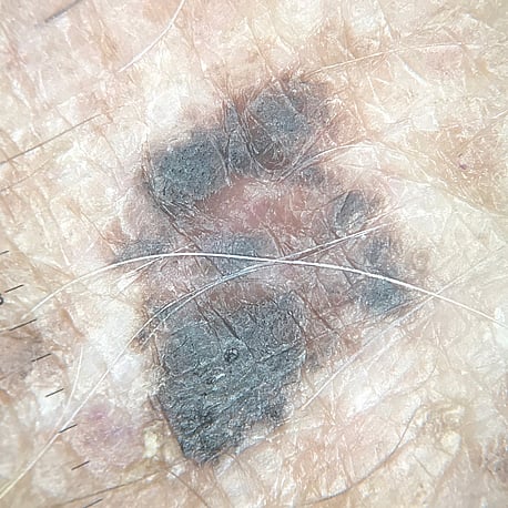
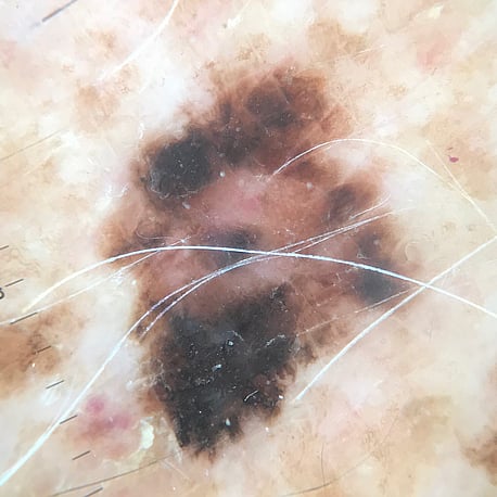
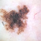
There are many benign lesions, lumps and bumps, but it’s hard to easily tell the difference between these and something of concern. If you have a new dark mole, or a spot that won’t heal, or something that’s just ‘odd-looking’ (the ugly duckling sign) this is a good reason to get a check. Any moles that you think are changing should be assessed. Has it changed shape and outline – say from a regular Rangitoto to a Waiheke Island? Rangitoto is an example of a mole that is mostly circular, uniform in colour and is smaller – whereas Waiheke is bigger, has differing colours and an irregular shape.
Read more on the main types of skin cancer HERE.
Rangitoto Lesion

Waiheke Lesion
Treatment
Accurate diagnosis by a skin cancer specialist will determine the most appropriate and effective treatment. I offer a variety of surgical and non-surgical treatments, and I will discuss what is best for you in every case.
Non-Surgical
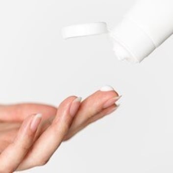
Topical treatments can be an effective non-surgical way to treat certain types of skin cancers and pre-cancerous lesions. They are quick and easy to use, and include applications of liquid nitrogen, or creams such as Efudex, and Imiquimod.
*Liquid nitrogen – This is a liquefied gas that boils at -196° Celsius. It is used to freeze and destroy superficial skin growths.
*Imiquimod (Aldara) – This cream is an immune response modifier. It causes the immune system to attack the growths it is applied to. This is often used for BCCs.
*5-Fluoruracil (Efudex) – This cream is a chemotherapy agent. It is effective against both pre-cancerous and cancerous cells, and has minimal effects on normal skin cells. This is more often used for solar keratosis and early/superficial SCCs.
Read more on the main types of skin cancer HERE
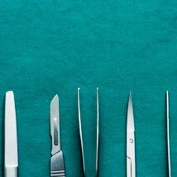
Surgical Treatment
Surgical treatments include biopsies - where the whole or part of a lesion is removed and tested. These can take the form of punch, shave or excision biopsies, depending on the lesion and its location. These are quick and simple to perform, and the tissue is checked by expert pathologists.
If a lesion needs removal, it can be safely and efficiently removed in our surgery. I am experienced in the use of advanced skin surgery including skin flaps and grafts, and get excellent results for my patients both surgically and cosmetically. Excision with clear margins is often the best way to eliminate skin cancer.
Ongoing management
Self Checks
Learn how to check your own skin and stay on top of changes. This should be done regularly, which is every 3–6 months for the general population, and in those at risk every 1–3 months.
Use a mirror, check between fingers and toes and don’t forget the scalp and soles of your feet. Does anything stand out? Assess lesions for changes in colour, size, thickness or outline Get lesions assessed if they are itchy, ulcerated or bleed. As a good guide - if something is an injury it should heal in three weeks. If the lesion you are watching doesn’t do that, get it checked.
Look out for ‘ugly ducklings’. These are lesions that stick out, and seem at odds with the others on your body. Any concerning lesions should be assessed by your GP or skin specialist. And remember you can check the skin of your family members too.